This is an artistic rendition of an immunofluorescent image of T cells in transplanted islets in mice. Foreign tissue such as transplanted islets are rejected without immunosuppression, which is a barrier for wider application of this therapy. Here, we used regulatory T cells (Tregs) to induce immune tolerance to the tissue so that immunosuppression is not needed. In this image, Tregs are those with green "rings" (cell membrane protein CD4) with "red stuffing" (transcription factor Foxp3). The blue rings are cell membrane protein CD8 that mark the killer T cells that reject the islets. In this image, they are kept at bay by the regulatory T cells. Without Treg therapy, the entire space would be filled with the killer T cells and islet would be gone. In this image, the protected islets are not visualized. Mouse studies such as this provided support to translate the therapy to humans. We are currently conducting a trial with the Tregs manufactured in the UCSF GMP facility (next image). Image courtesy of Karim Lee, PhD, a former postdoc in Tang Lab.
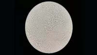
Human Treg in culture: at the UCSF GMP facility, we produce clinical grade Tregs to support 10 early phase clinical trials. During the production, we purify Tregs from blood and grow them for 2 weeks to increase their number and function before infusing to patient. This image shows a very happy Treg culture in the middle of cell manufacture. Image courtesy of Yani Peng, Tang Lab.
The image shows a pancreatic islet stained with a red dye that labels the outer leaflet of cell membrane so that the cellular organization within an islet can be visualized. The yellow green color marks macrophages that naturally reside in islets. the image is a thin optical section in the mid-section of the islet captured using a confocal microscope (by me!). Islet transplantation is a curative therapy for people with type 1 diabetes. Image courtesy Tang Lab.
Superresolution image of a group of killer T cells (green and red) surrounding a cancer cell (blue, center). When a killer T cell makes contact with a target cell, the killer cell attaches and spreads over the dangerous target. The killer cell then uses special chemicals housed in vesicles (red) to deliver the killing blow. This event has thus been nicknamed “the kiss of death”. After the target cell is killed, the killer T cells move on to find the next victim. Image courtesy of Alex Ritter, Jennifer Lippincott Schwartz and Gillian Griffiths, National Institutes of Health
A new 44,000-square-foot, state-of-the-art cell therapy development, manufacturing and collaboration center will be located in leased space on UCSF’s Mission Bay campus.
Photo by Matt Beardsley
Foundational toolkit for programming cell function.
Image courtesy the Cell Design Institute https://www.celldesigninstitute.org/
Cell engineering infographic.
Image courtesy the Cell Design Institute https://www.celldesigninstitute.org/


